Why would I need a breast biopsy?
If an anomaly (e.g., a lump, skin thickening, skin texture change, nipple color change, or evidence of nipple discharge) is discovered during a breast self-examination or routine screening, your physician will schedule a breast biopsy to test for cancer or other potentially serious medical concerns. Biopsies are the only definitive way to confirm whether breast tissue is benign or cancerous.
Your doctor may require a breast biopsy after a diagnostic mammogram, breast MRI, or breast ultrasound shows an unusual growth or mass within the breast.
Needle Biopsy
Biopsies are the only definitive way to confirm whether breast tissue is benign or cancerous.
During a needle biopsy, small amounts of breast tissue from an abnormal lesion are removed through a needle and then studied under a microscope by a pathologist, who provides a diagnosis. A needle biopsy is performed with a local anesthetic and minimal discomfort. No stitches are required, and most women can resume normal daily activities immediately.
If you have discovered an abnormality in your breast, the next diagnostic step is likely an ultrasound breast biopsy. This biopsy extracts a portion of suspicious tissue for analysis by a pathologist.
Ultrasound technology allows your healthcare provider to quickly and accurately identify changes that might indicate the development of serious illness. Ultrasound tools use soundwave energy to generate images of internal anatomical structures and eliminate the need to expose patients to radiation from X-ray, computed tomography (CT scan), or mammography technology.
Here’s what you need to know about having an ultrasound breast biopsy.
Ultrasound Breast Biopsy
If your mammogram or ultrasound exam shows an area of concern, an ultrasound-guided biopsy may be recommended. Valley Radiology's specialized breast radiologists use ultrasound to guide them while taking small samples of tissue from the area of concern for analysis by a pathologist.
Some common reasons for this procedure are:
- A suspicious solid mass
- A distortion in the structure of the breast tissue
- An area of abnormal tissue changes
Stereotactic Breast Biopsy
A stereotactic breast biopsy is performed when a mammogram shows a breast abnormality such as:
- A suspicious solid mass
- Microcalcifications (a tiny cluster of small calcium deposits)
- A distortion in the structure of the breast tissue
- An area of abnormal tissue change
- A new mass or area of calcium deposits present at a previous surgery site
How is a biopsy procedure performed?
A breast biopsy allows your medical team to identify potentially dangerous changes to your breast tissue early and with minimal discomfort. A specially trained radiologist will perform your breast biopsy.
A breast biopsy might be conducted in one of several ways. One of the most common procedures is an ultrasound-guided core needle breast biopsy. This technique is most often used when a breast abnormality is visibly on ultrasound. A core needle is a hollow needle into which tissue and fluid samples can be collected.
During this procedure, the patient will lie on their back or turn slightly to the side. Local anesthetic is injected to ensure comfort. The doctor or breast sonographer runs a handheld ultrasound device over the breast until the suspicious mass becomes visible on a computer screen. The doctor then makes a small incision near the mass through which the biopsy needle can be inserted into the mass, and tissue samples are then withdrawn for evaluation. Typically, several samples are extracted.
The radiologist will then place a small clip called a tissue marker in the breast to mark the place where the tissue was removed. After the clip is placed, the breast will be placed in compression for approximately 10 minutes to stop any bleeding. The nurse will then place small steristrips (like tape) over the incision. No stitches are needed.
A mammogram may be performed to ensure that the marker is in the correct position.
Another type of breast biopsy technique is a stereotactic core needle or vacuum-assisted device biopsy. A specially trained radiologist will perform your stereotactic breast biopsy. The affected breast or breasts will be compressed and held in position throughout the procedure. A local anesthetic will be injected into the breast to numb it.
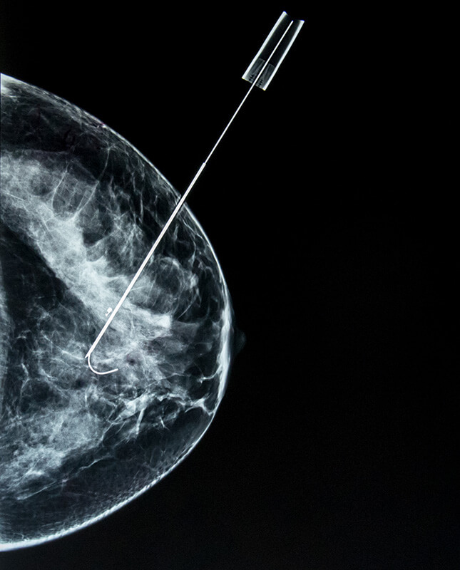
Several stereotactic pairs of X-ray images will be taken. A tiny nick will be made in the skin at the site where the biopsy needle is inserted. The radiologist will then insert the needle and advance it to the location of the abnormality using the X-ray and computer-generated coordinates. X-ray images are again obtained to confirm that the needle tip is actually within the lesion.
Tissue samples are removed using one of two methods:
- In a core needle biopsy, the automated mechanism is activated, moving the needle forward to obtain “cores” of breast tissue. The outer sheath instantly moves forward to cut the tissue and keep it in the trough. This process is repeated three to six times.
- With a vacuum-assisted device (VAD), vacuum pressure is used to pull tissue from the breast through the needle into the sampling chamber. Without withdrawing and reinserting the needle, it rotates positions and collects additional samples. Typically, eight to 10 samples of tissue are collected from around the lesion.
After the sampling, the needle will be removed and a final set of images will be taken.
A small tissue marker may be placed at the site for future localization if needed. Once the biopsy is complete, pressure will be applied to stop any bleeding, and the opening in the skin will be covered with a dressing. No sutures are required. A mammogram may be performed to ensure that the marker is in the correct position. This procedure typically takes about an hour to complete.
How do I prepare for a breast biopsy?
Please discuss the following with our breast team:
- If you have allergies
- If you have taken aspirin within the week of the scheduled procedure
- If you are currently taking any blood-thinning medication
- If you are pregnant or think you could be pregnant
Your doctor will provide you with specific instructions prior to your procedure.
How long does a breast biopsy take?
A breast biopsy takes approximately one hour. Your doctor will give you a detailed breakdown of your procedure during your consultation.
What are the risks of a breast biopsy?
Ultrasound-guided and Stereotactic breast biopsies are minimally invasive procedures and do not carry the same risks of complications as open surgeries. However, there is a minor infection risk at the biopsy site, so it’s important to follow your doctor’s aftercare instructions closely.
Common issues experienced after a breast biopsy include:
- Minor bruising
- Inflammation
- Redness
It isn’t unusual to experience discomfort after the procedure. Ice packs and over-the-counter acetaminophen medications like Tylenol are effective for reducing pain.
Both patients and physicians prefer ultrasound-guided breast biopsies to traditional surgical biopsies because they are significantly less painful and more efficient. They also offer the following benefits:
- Less damage to breast tissue than surgical biopsy
- No radiation exposure
- Effective for visualizing and retrieving tissue growths in difficult-to-access regions, like under the arm
Please note that a doctor's referral is required for a breast biopsy. However, for annual screening mammograms, no physician's referral is required. Breast experts still agree that 40 is the age to start your annual screening. Please make a point to schedule screening mammograms every year and urge all of the women in your life to do the same. We offer flexible scheduling at Schedule Your Mammogram.