Week One
VALLEY RADIOLOGY Interventional Radiology Case of the Week:
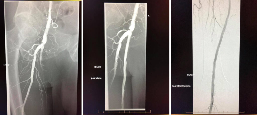
Case of the week: 70 year old patient with severe right leg pain with walking. Angiogram shows complete occlusion of the right superficial femoral artery. We were able to cross the occlusion with catheter and reconstruct the artery with balloon angioplasty and stents! All done without the need for open surgery!
Week Two
VALLEY RADIOLOGY Interventional Radiology Case of the Week:
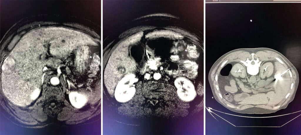
Case of the week: 56 year old with multi focal Hepatocellular Carcinoma with 3 liver masses. Staged treatment with CT guided microwave ablation followed by intra-arterial chemoembolization (chemotherapy coated beads injected directly into the blood supply of the liver). Great stuff Dr Meka!!
Week Three
VALLEY RADIOLOGY Interventional Radiology Case of the Week:
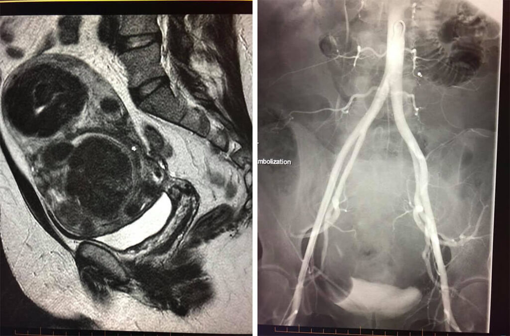
38 year old female with large fibroids in the uterus. Uterus is size of 6 month pregnancy. She complains of severe pain and irregular bleeding because of the fibroids. She elected to have an uterine artery embolization (also known as uterine fibroid embolization UAE/UFE) instead of surgery. We inserted a microcatheter into the uterine arteries and blocked off the blood supply to the fibroids with microspheres. 1 month later all her symptoms have resolved. 6 months later her fibroids have all shrunk or disappeared with no blood flow to the fibroids. And uterine function is normal!!
Week Four
INTERVENTIONAL RADIOLOGY by Valley Radiology
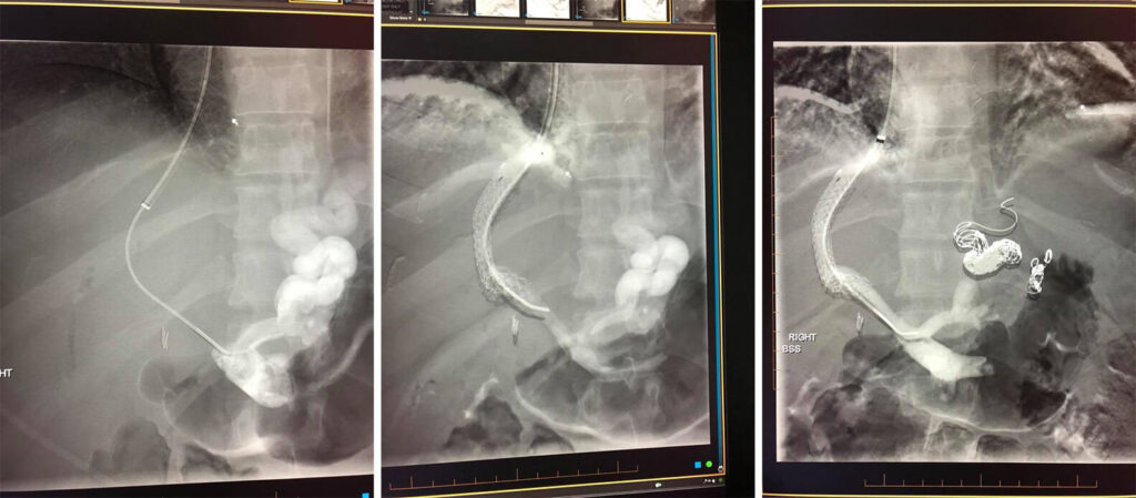
62 yo male with cirrhosis and massive GI bleeding. Treated in ER with endoscopy and blind banding. Thanks Dr Joseph Henderson of Fayetteville Gastroenterology Associates! Patient was then transferred to IR suite for an emergency TIPS (transjugular intrahepatic portosytemic shunt) and embolization of the esophageal and gastric varices.
Week Five
INTERVENTIONAL RADIOLOGY by Valley Radiology
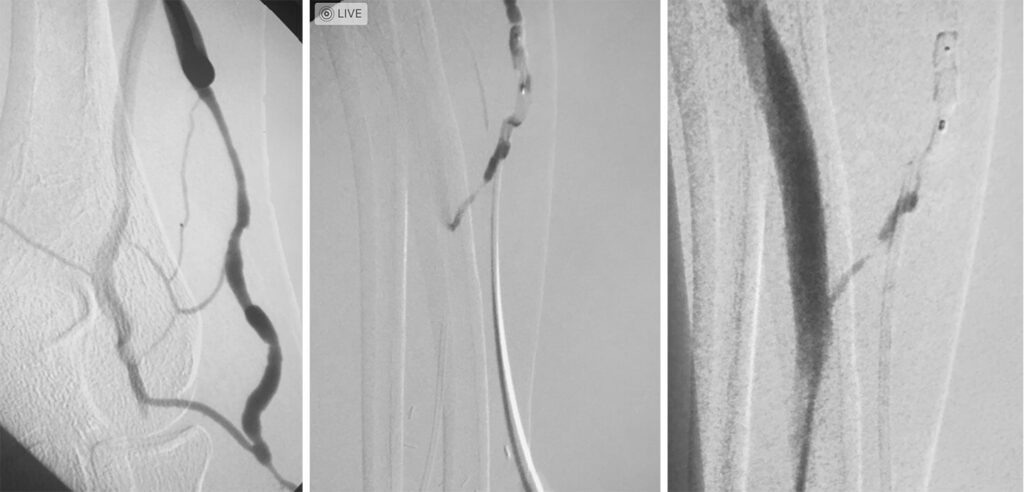
Case of the week: Diaylsis patient with decrease in functioning of AV fistula - competing accessory vein needs to be shut down to fully route flow to AV fistula
Plug embolization of a competing vein for a successful maturation of the left arm AV fistula.
(*closing off of larger vein that takes blood away from a normal AV fistula)
Photo 1 - Accessory vein to shut down
Photo 2- Plug insertion into competing accessory vein
Photo 3- Full functioning AV fistula due to plug embolization
Week Six
VALLEY RADIOLOGY Interventional Radiology Case of the Week:
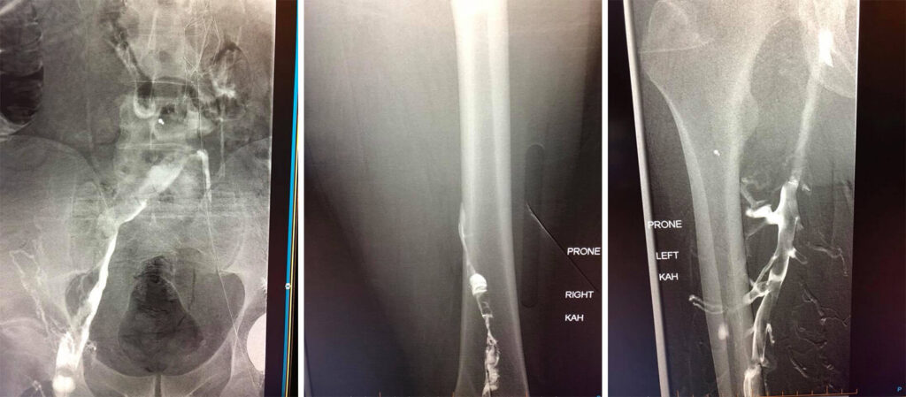
Case of the week: A 60 year old gentleman with extensive blood clots throughout both legs, pelvis and abdomen. Patient has an IVC filter with clot extending about the filter and acute clots into the lungs.
Day 1 venogram pictures shows occlusive deep vein thrombosis without any venous flow through the legs, pelvis or abdomen. Catheters were placed to drip clot busting drugs directly into the clot for 24 hours.
Day 2 venogram shows improved flow through the veins. Residual clot was then removed with special suction catheter (thrombectomy).
Week Seven
VALLEY RADIOLOGY Interventional Radiology Case of the Week:
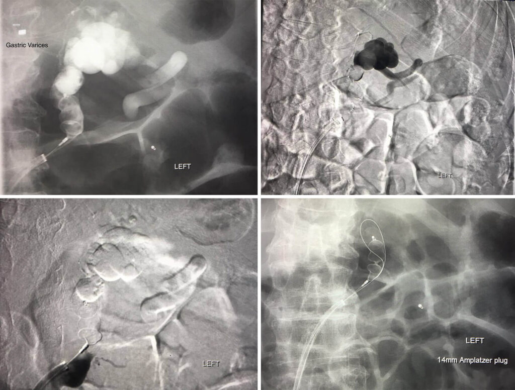
Plug assisted Retrograde Transveonous Obliteration of the Gastric Varices.
The Interventional approach to treating an upper GI bleed!!!!
76 year old male patient with Upper GI bleed. Endoscopy showed gastric varices.
Patient sent to Interventional Radiology to treat the varices with a minimal invasive approach through the common femoral vein in the groin. Catheter then utilized to cannulate left renal vein then retracted to select the adrenal vein/splenorenal shunt to get to the gastric varices. Amplatzer plug placed to stop the bleeding. Great work Dr. Meka!!!
Week Eight
VALLEY RADIOLOGY Interventional Radiology Case of the Week:
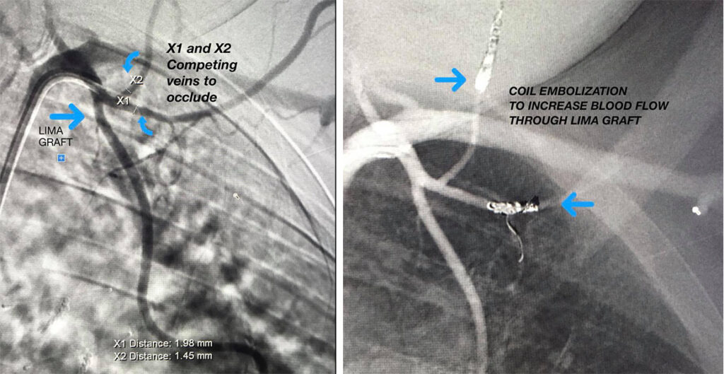
Patient with recurrent chest pain. Patient has had a LIMA Graft placed previously. LIMA- Left Internal Mammary Artery used for CABG - Coronary Artery bypass grafting. Competing arteries can take away blood flow through the LIMA graft. Dr. Meka received this patient and used coil embolization to block off the competing arteries (X1 and X2) and revascularize the LIMA graft by occluding the competing arteries.
